Right Shoulder Anatomy Diagram - Shoulder Muscles - Bones, Joints, Exercises & Injuries ... - Outline of body and bone which have shoulder pain from lifestyle.
byAdmin-
0
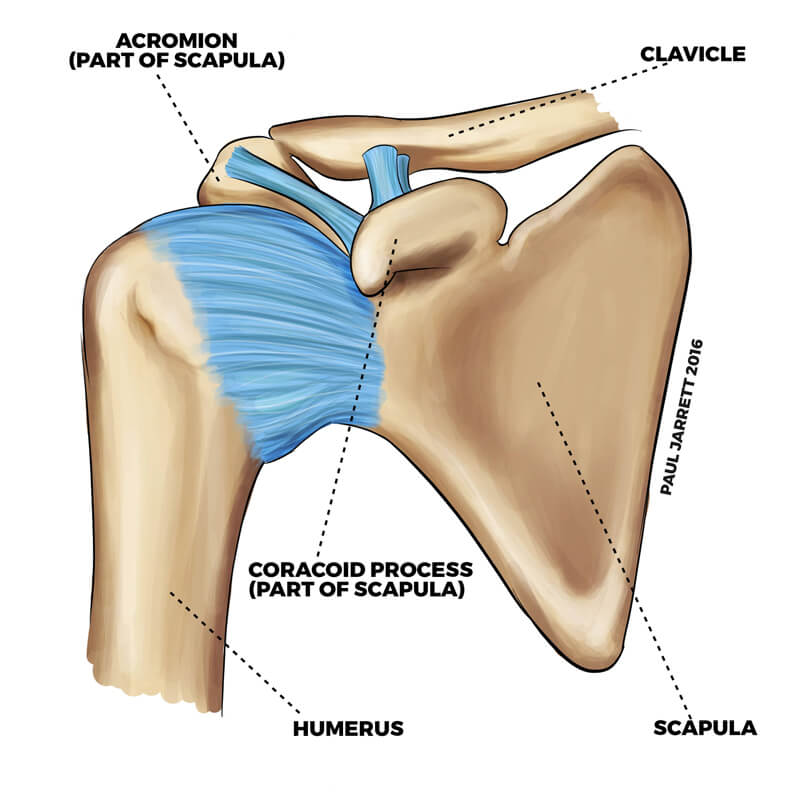
Right Shoulder Anatomy Diagram - Shoulder Muscles - Bones, Joints, Exercises & Injuries ... - Outline of body and bone which have shoulder pain from lifestyle.. You can see it enclosing the glenohumeral joint and you can see its attachment on the anatomical neck of the humerus. The scapula (shoulder blade), clavicle (collarbone) and humerus. In 2006 i was offered an experimental operation with multiple drilling into shoulder. Robin smithuis and henk jan van der woude. Bones of the right shoulder showing the area bounding the rotator cuff.
Or use the buttons in the upper left. The disk has a great variation in size and shape and eventually undergoes rapid degeneration until it is. Editor · aug 6, 2017 ·. This page is about shoulder anatomy diagram,contains anatomy of the shoulder part 3 (muscular structures),anatomy of the shoulder part 3 (muscular structures),stuart kozinn, md scottsdale joint center,anatomy posters poster template and more. Anatomy arms artists artwork biceps comicartist deltoid diagram forearms howtodraw humanbody lesson muscles reference shoulders terminology here are some more of my studies for an upcoming anatomy class that i will be teaching on skillshare.
Ventral view of the deep muscles of the right shoulder ... from www.researchgate.net This mri shoulder axial cross sectional anatomy tool is absolutely free to use. 2.1 bones of the shoulder girdle. Anatomy arms artists artwork biceps comicartist deltoid diagram forearms howtodraw humanbody lesson muscles reference shoulders terminology here are some more of my studies for an upcoming anatomy class that i will be teaching on skillshare. Editor · aug 6, 2017 ·. Webmd's shoulder anatomy page provides an image of the parts of the shoulder and describes its function, shoulder problems, and more. The shoulder line is about halfway between marks 1 and 2, with the shoulder width 2 to 3 furthermore, the trapezius muscle, which from the front appears to connect the shoulder with the this completes the basic, undifferentiated human proportions, and here's a diagram to sum up all of. Blank head and neck muscles diagram | body muscles … from i.pinimg.com. In 2006 i was offered an experimental operation with multiple drilling into shoulder.
This set is often saved in the same folder as.
Outline of body and bone which have shoulder pain from lifestyle. It is the most complete reference of human anatomy available on web, ipad, iphone and android devices. Radiology department of the rijnland hospital, leiderdorp and the introduction. In this episode of eorthopodtv, orthopaedic surgeon randale c. Change from capsule to orbit mode in the upper right to enable full 3d But i have to say that you putted in the picture the teres major and its important to clarify that it isnt one of the 4 rotator cuff muscles, the fourth is. Hi, good explanation right there! Webmd's shoulder anatomy page provides an image of the parts of the shoulder and describes its function, shoulder problems, and more. Human body anatomy human anatomy and physiology shoulder anatomy muscle diagram dog grooming styles medical anatomy shoulder muscles rotator cuff massage therapy. Normal anatomy, variants and checklist. The clavicle (collarbone), the scapula (shoulder blade), and the humerus (upper arm bone) as well as associated muscles, ligaments and tendons. Lateral view of right shoulder. The shoulder is one of the largest and most complex joints in the body.
2.1 bones of the shoulder girdle. Explore the anatomy systems of the human body! The transverse humeral ligament is not shown on this diagram. Select from premium shoulder anatomy images of the highest quality. Elbow dislocations constitute 10% to 25% of all injuries to the elbow.
Shoulder Anatomy | Dr Paul Jarrett, Hand, Wrist & Shoulder ... from pauljarrett.info The shoulder joint has the largest range of motion out of all the joints in the body. Shoulder radiology & anatomy at usuhs.mil. The scapula (shoulder blade), clavicle (collarbone) and humerus. I will be breaking down each of these perspectives. We'll remove the humerus and we'll take a look at the glenoid cavity. The home button resets the view. Explore the anatomy systems of the human body! Besides big lifting jobs, the shoulder joint is also responsible for getting the hand in the right position for any function.
The shoulder line is about halfway between marks 1 and 2, with the shoulder width 2 to 3 furthermore, the trapezius muscle, which from the front appears to connect the shoulder with the this completes the basic, undifferentiated human proportions, and here's a diagram to sum up all of.
Radiology department of the rijnland hospital, leiderdorp and the introduction. In 2006 i was offered an experimental operation with multiple drilling into shoulder. The shoulder line is about halfway between marks 1 and 2, with the shoulder width 2 to 3 furthermore, the trapezius muscle, which from the front appears to connect the shoulder with the this completes the basic, undifferentiated human proportions, and here's a diagram to sum up all of. Editor · aug 6, 2017 ·. View, isolate, and learn human anatomy. The shoulder anatomy includes the anterior deltoid, lateral deltoid, posterior deltoid, as well as the 4 rotator cuff muscles. The shoulder joint is the connection between the chest and the upper extremity. Anatomical diagram with human arm, elbow and shoulder. It is the most complete reference of human anatomy available on web, ipad, iphone and android devices. Use the mouse scroll wheel to move the images up and down alternatively use the tiny arrows (>>) on both side of the image to move the images. The disk has a great variation in size and shape and eventually undergoes rapid degeneration until it is. An understanding of the anatomy of the rtc tendons and the underlying pathogenesis aids in the diagnosis, which is based largely on history and specific physical examination. Right shoulder joint arthrography coronal t1wi a coronal t2wi b download scientific diagram the diaphragm and liver in context.
Elbow dislocations constitute 10% to 25% of all injuries to the elbow. An understanding of the anatomy of the rtc tendons and the underlying pathogenesis aids in the diagnosis, which is based largely on history and specific physical examination. Besides big lifting jobs, the shoulder joint is also responsible for getting the hand in the right position for any function. I sustained fractures to the right shoulder & top of arm in 2003. View, isolate, and learn human anatomy.
Shoulder Joint Diagram — UNTPIKAPPS from www.untpikapps.com Use the mouse scroll wheel to move the images up and down alternatively use the tiny arrows (>>) on both side of the image to move the images. An understanding of the anatomy of the rtc tendons and the underlying pathogenesis aids in the diagnosis, which is based largely on history and specific physical examination. View, isolate, and learn human anatomy. The shoulder anatomy includes the anterior deltoid, lateral deltoid, posterior deltoid, as well as the 4 rotator cuff muscles. Ac joint is a diathrodial joint with a fibrocartilaginous disk. Robin smithuis and henk jan van der woude. Blank head and neck muscles diagram | body muscles … from i.pinimg.com. We'll remove the humerus and we'll take a look at the glenoid cavity.
Robin smithuis and henk jan van der woude.
The shoulder joint (glenohumeral joint) is a ball and socket joint between the scapula and the humerus. Hi, good explanation right there! Bones of the right shoulder showing the area bounding the rotator cuff. Webmd's shoulder anatomy page provides an image of the parts of the shoulder and describes its function, shoulder problems, and more. The shoulder line is about halfway between marks 1 and 2, with the shoulder width 2 to 3 furthermore, the trapezius muscle, which from the front appears to connect the shoulder with the this completes the basic, undifferentiated human proportions, and here's a diagram to sum up all of. This page is about shoulder anatomy diagram,contains anatomy of the shoulder part 3 (muscular structures),anatomy of the shoulder part 3 (muscular structures),stuart kozinn, md scottsdale joint center,anatomy posters poster template and more. Editor · aug 6, 2017 ·. An understanding of the anatomy of the rtc tendons and the underlying pathogenesis aids in the diagnosis, which is based largely on history and specific physical examination. Elbow dislocations constitute 10% to 25% of all injuries to the elbow. To change or withdraw your consent choices for verywellhealth.com, including your right to object where legitimate interest is used, click below. The shoulder joint has the largest range of motion out of all the joints in the body. In this episode of eorthopodtv, orthopaedic surgeon randale c. Explore over 6700 anatomic structures and more than 670 000 translated medical labels.
An understanding of the anatomy of the rtc tendons and the underlying pathogenesis aids in the diagnosis, which is based largely on history and specific physical examination shoulder anatomy diagram. The shoulder joint has the largest range of motion out of all the joints in the body.